Zhodnocení vztahu mezi distribuční šíří objemu erytrocytů (anisocytózou) a stupněm jaterní fibrózy u dětí s chronickými jaterními chorobami
Seyed Mohsen Dehghani1, Masoud Tahani2, Mahammad Reza Bordbar1, Mahsa Borjzadeh Gashtaseb1, Iraj Shahramian1
+ Pracoviště
Souhrn
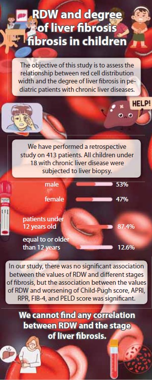
Úvod: Distribuční šíře objemu erytrocytů (RDW) popisuje různorodost v objemech erytrocytů a je součástí celkového krevního obrazu. Nedávné studie ovšem ukázaly na vztah mezi RDW a zvýšenou úmrtností u mnoha klinických stavů a zjistily, že vysoký RDW navyšuje pravděpodobnost úmrtí z jakýchkoli příčin. Některé studie také popisují vztah mezi hodnotami RDW a závažností jaterních onemocnění. Byla popsána přímá úměrnost mezi hodnotami RDW a MELD skóre u různých stadií infekce virem hepatitidy B. Zároveň s tím se hodnota RDW zvyšovala úměrně zhoršujícímu se stupni Child-Pugh skóre jaterní cirhózy. Metoda: Tato studie zkoumala klinické využití hodnot RDW pro určování přítomnosti jaterní fibrózy u dětí s chronickým onemocněním jater. Provedli jsme retrospektivní studii zahrnující 413 pacientů. Posbírali jsme demografická, klinická a laboratorní data a histologické nálezy stadií fibrózy z lékařských záznamů a analyzovali jsme je pomocí SPSS. Výsledky: Naše studie neukázala významnou korelaci mezi hodnotami RDW a stupněm jaterní fibrózy. Naproti tomu asociace mezi RDW a zhoršujícím se Child-Pugh skóre, APRI, RPR, FIB-4, a PELD skóre významná je. Závěr: Nenašli jsme žádný vztah mezi hodnotami RDW a stadiem jaterní fibrózy.
Klíčová slova
cirhóza, játra, fibróza, RDW
Článek je v angličtině, prosím přepněte do originální verze.
Pro přístup k článku se, prosím, registrujte.
Výhody pro předplatitele
Výhody pro přihlášené
Literatura
1. Hudacko R, Theise N. Liver biopsies in chronic viral hepatitis: beyond grading and staging. Archives of pathology & laboratory medicine. Arch Pathol Lab Med 2011; 135(10): 1320–1328. doi: 10.5858/arpa.2011-0021-RA.
2. Ghany MG, Strader DB, Thomas DL et al. Diag- nosis, management, and treatment of hepatitis C: an update. Hepatology 2009; 49(4): 1335–1374. doi: 10.1002/hep.22759.
3. Lok AS, McMahon BJ. Chronic hepatitis B. Hepatology 2007; 45(2): 507–539. doi: 10.1002/hep. 21513.
4. Czaja AJ. Performance parameters of the conventional serological markers for autoimmune hepatitis. Digestive diseases and sciences. Dig Dis Sci 2011; 56(2): 545–554. doi: 10.1007/s10620-010-1501-1.
5. Abdollahi MR, Somi MH, Faraji E. Role of international criteria in the diagnosis of autoimmune hepatitis. World J Gastroenterol 2013; 19(23): 3629–3633. doi: 10.3748/wjg.v19.i23.3629.
6. Berasain C, Betes M, Panizo A et al. Pathological and virological findings in patients with persistent hypertransaminasaemia of unknown aetiology. Gut 2000; 47(3): 429–435. doi: 10.1136/gut.47.3.429.
7. Hano H, Takasaki S. Three-dimensional observations on the alterations of lobular architecture in chronic hepatitis with special reference to its angioarchitecture for a better understanding of the formal pathogenesis of liver cirrhosis. Virchows Arch 2003; 443(5): 655–663. doi: 10.1007/s00428-003-0843-x.
8. Degos F, Perez P, Roche B et al. Diagnostic accuracy of FibroScan and comparison to liver fibrosis biomarkers in chronic viral hepatitis: a multicenter prospective study (the FIBROSTIC study). J Hepatol 2010; 53(6): 1013–1021. doi: 10.1016/j.jhep.2010.05.035.
9. Poynard T, Imbert-Bismut F, Munteanu M et al. Overview of the diagnostic value of biochemical markers of liver fibrosis (FibroTest, HCV FibroSure) and necrosis (ActiTest) in patients with chronic hepatitis C. Comp Hepatol 2004; 3(1): 8. doi: 10.1186/1476-5926-3-8.
10. Cadranel JF, Rufat P, Degos F. Practices of liver biopsy in France: results of a prospective nationwide survey. For the Group of Epidemiology of the French Association for the Study of the Liver (AFEF). Hepatology 2000; 32(3): 477–481. doi: 10.1053/jhep.2000.16602.
11. Kleinman RE. Walker’s pediatric gastrointestinal disease. USA: PMPH 2018.
12. Heimbach JK, Kulik LM, Finn RS et al. AASLD guidelines for the treatment of hepatocellular carcinoma. Hepatology 2018; 67(1): 358–380. doi: 10.1002/hep.29086.
13. Yeung CY, Lee HC, Chan WT et al. Vertical transmission of hepatitis C virus: Current knowledge and perspectives. World J Hepatol 2014; 6(9): 643–651. doi: 10.4254/wjh.v6.i9.643.
14. Tian L, Ye Z, Kafka K et al. Biliary atresia relevant human induced pluripotent stem cells recapitulate key disease features in a dish. J Pediatr Gastroenterol Nutr 2019; 68(1): 56–63. doi: 10.1097/MPG.0000000000002187.
15. Perlmutter DH. Liver injury in alpha 1-antitrypsin deficiency: an aggregated protein induces mitochondrial injury. J Clin Invest 2002; 110(11): 1579–1583. doi: 10.1172/JCI16787.
16. Weiss G, Goodnough LT. Anemia of chronic disease. N Engl J Med 2005; 352(10): 1011–1023. doi: 10.1056/NEJMra041809.
17. Weiss G, Ganz T, Goodnough LT. Anemia of inflammation. Blood 2019; 133(1): 40–50. doi: 10.1182/blood-2018-06-856500.
18. Kliegman RM, Behrman RE, Jenson HB et al. Nelson textbook of pediatrics e-book. Elsevier Health Sciences 2016.
19. Wyllie R, Hyams JS. Pediatric Gastrointestinal and Liver Disease E-Book: Expert Consult-Online and Print. Elsevier Health Sciences 2016.
20. Kasper DL, Fauci AS, Hauser SL et al. Harrison’s principles of internal medicine. McGraw Hill Education 2018: 379–387.
21. Lippi G, Salvagno GL, Guidi GC. Red blood cell distribution width is significantly associated with aging and gender. Clin Chem Lab Med 2014; 52(9): e197–199. doi: 10.1515/cclm-2014-0353.
22. Xu W-S, Qiu X-M, Ou Q-s et al. Red blood cell distribution width levels correlate with liver fibrosis and inflammation: a noninvasive serum marker panel to predict the severity of fibrosis and inflammation in patients with hepatitis B. Medicine (Baltimore) 2015; 94(10): e612. doi: 10.1097/MD.0000000000000612.
23. Chen B, Ye B, Zhang J et al. RDW to platelet ratio: a novel noninvasive index for predicting hepatic fibrosis and cirrhosis in chronic hepatitis B. PLoS One 2013; 8(7): e68780. doi: 10.1371/journal.pone.0068780.
24. Huang R, Yang C, Wu K et al. Red cell distribution width as a potential index to assess the severity of hepatitis B virus‐related liver diseases. Hepatol Res 2014; 44(14): E464–E470. doi: 10.1111/hepr.12342.
25. Ishak K, Baptista A, Bianchi et al. Histological grading and staging of chronic hepatitis. J Hepatol 1995; 22(6): 696–699. doi: 10.1016/0168-8278(95)80226-6.
26. Berger D, Desai V, Janardhan S. Con: liver biopsy remains the gold standard to evaluate fibrosis in patients with nonalcoholic fatty liver disease. Clin Liver Dis (Hoboken) 2019; 13(4): 114–116. doi: 10.1002/cld.740.
27. Mahjoubin-Tehran M, De Vincentis A, Mikhailidis DP et al. Non-alcoholic fatty liver disease and steatohepatitis: State of the art on effective therapeutics based on the gold standard method for diagnosis. Mol Metab 2021; 50: 101049. doi: 10.1016/j.molmet.2020.101049.
28. Hu Z, Sun Y, Wang Q et al. Red blood cell distribution width is a potential prognostic index for liver disease. Clin Chem Lab Med 2013; 51(7): 1403–1408. doi: 10.1515/cclm-2012-0704.
29. Abdollahi M, Pouri A, Ghojazadeh M et al. Noninvasive serum fibrosis markers: A study in chronic hepatitis. BioImpacts 2015; 5(1): 17–23. doi: 10.15171/bi.2015.05.